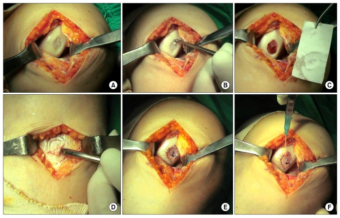
Fig. 2.
Surgical procedure of the 1st generation autologous chondrocyte implantation. (A) Outerbridge 4 lesion in the medial femoral condyle. (B) Debridement of the calcified cartilage layer and unstable chondral flap. (C) Defect size is measured with a sterile paper. A 2 mm oversized template is needed. (D) Periosteal flap is excised from the proximal medial tibia. (E) The periosteal flap watertightly covers the defect. (F) Chondrocyte suspension is injected to the defect through a plastic 18-gauge angiocath needle.

