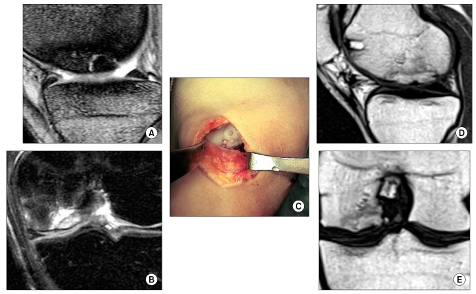
Fig. 6.
Osteochondral plug fixation for the treatment of osteochondritis dissecans (OCD). (A) Sagittal T2 fast spin echo (FSE) image showing stage 3 OCD lesion in the medial femoral condyle. The lesion is partially separated. (B) Coronal T2 fat suppression FSE image showing OCD lesion with focal bone marrow edema. (C) Fixation of OCD with multiple osteochondral plugs. (D) Sagittal image. (E) Coronal image showing the OCD lesions completely incorporated to the host bone at 24 months after surgery.

