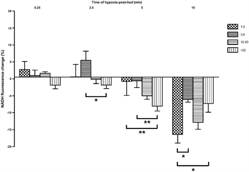
Fig. 7.

Change in NADH fluorescence (%) in hippocampal slices from 1–2, 3–6, 12–20, >22 months old rats following varying lengths of hypoxia post-hsd (0.25, 2.5, 5 and 10 min). Following 2.5 min hypoxia post-hsd, NADH hyperoxidation was significantly greater in slices from >22 months old rats (n = 9) compared to slices from 3 to 6 months old rats (n = 7; *p < 0.05). This effect was amplified between these age groups at 5 min hypoxia (**p < 0.01). In addition, the percentage of hyperoxidation was greater following 5 min hypoxia in slices from >22 months old rats (n = 6) compared to 1–2 months old rats (n = 7; **p < 0.01). Ten minutes of hypoxia post-hsd resulted in the greatest amount of hyperoxidation in all age groups with slices from 1 to 2 months old rats (n = 4) exhibiting significantly higher amounts of hyperoxidation compared to slices from 3 to 6 months old rats (n = 4) and >22 months old rats (n = 6; *p < 0.05).
