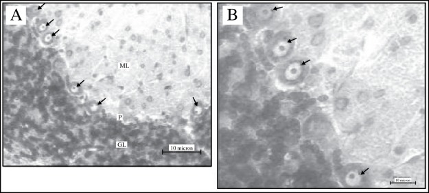
Figure 2 :
Photomicrographs of the cerebellar cortex of the deltamethrin-treated rat which had been stained with cresyl fast violet at lower magnification (A) (20x objective) and at higher magnification (B) (40x objectives). The Purkinje cells (arrows) lie in a monolayer between the molecular (ML) and granular layer (GL). Note the prominent nucleolus in each of the Purkinje cell.

