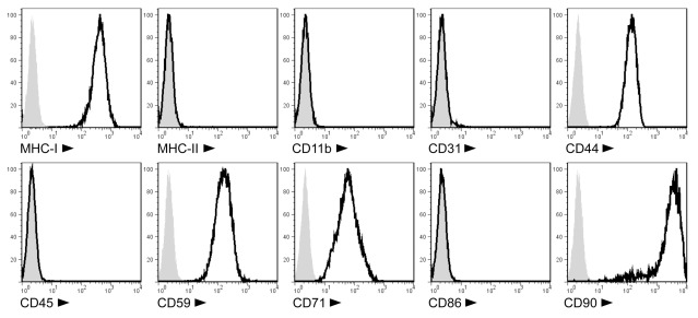
Figure 1.
Cell surface phenotype of PVG.7B MSC isolated from rat BM. Ex vivo-expanded MSC express MHC class I (RT1-A), CD44H (H-CAM), CD59 (MAC inhibitor), CD71 (transferrin receptor), and CD90 (Thy-1), but not MHC class II (RT1-B/D), CD11b (MAC-1), CD31 (PECAM-1), CD45 (LCA), or CD86 (B7-2) as detected by single-color flow cytometry. Representative stainings of MSC derived from PVG.7B BM are displayed as histograms [x-axis, signal intensity (log); y-axis, relative cell count (percent of max)] of surface antigens (solid line) and negative controls (gray histograms).

