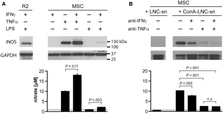
Figure 5.
Tumor necrosis factor α and IFNγ synergistically induce iNOS expression and NO production by MSC. 1 × 10⁴ MSC (PVG.7B) were stimulated for 24 h with combinations of IFNγ, TNFα, and a TLR agonist (A) or by medium supernatants from ConA-activated LNC (PVG.7B or PVG.1U) cultures (B). Pooled triplicates and quadruplicates, respectively, were analyzed for iNOS expression by Western blot with GAPDH assayed as a loading control. Nitrate concentrations were determined in the culture supernatants of the same wells (bottom panels). (A) MSC produced iNOS (130 kDa) in response to TNFα (25 ng mL−1) alone or together with IFNγ (100 U mL−1) or by a combination of IFNγ and LPS (100 ng mL−1). Nitrate concentration levels correlated with the observed levels of iNOS protein expression. The dotted line indicates baseline NO production without cytokines added. The R2 macrophage cell line was used as positive control for iNOS expression. (B) Supernatants from ConA-stimulated (ConA–LNC-sn) and unstimulated LNC cultures as a control (LNC-sn; baseline) were added to MSC cultures. Neutralizing antibodies (each 10 μg mL-1 final concentration) against either IFNγ or TNFα or both (indicated) were added at the start of culture and incubated for 24 h. iNOS was induced by addition of supernatant from stimulated LNC cultures and was blocked by IFNγ-specific antibody and, more potently, by TNFα-specific antibody alone or in combination. Nitrate concentrations correlated with the observed iNOS expression levels. Data are representative of four independent experiments. Nitrate concentration data are shown as the mean and the SD of triplicates or quadruplicates.

