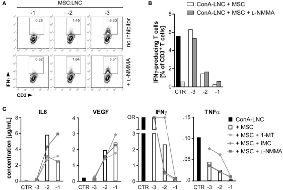
Figure 6.
MSC inhibition of IFNγ and TNFα secretion is not dependent on iNOS activity, but on COX-dependent production of prostaglandin. (A,B) MSC (PVG.7B) were added at the indicated dilutions [log (MSC:LNC)] at the start of ConA–LNC (PVG.7B) cultures. Cells from triplicate wells were pooled after 24 h and the number of IFNγ-positive T cells (IFNγ+CD3+ lymphocyte gate, values indicate relative frequencies) determined by flow cytometry. Inhibition of iNOS by L-NMMA (bottom row and shaded bars) did not influence the MSC-mediated suppression of IFNγ expression (top row and unshaded bars). Controls in (B) show IFNγ production by T cells alone in the presence (black) or absence (light gray) of ConA. (C) MSC (PVG.1U) were added at the indicated cell ratios [log] at the start of ConA–LNC (PVG.7B) cultures in the absence (white bars) or presence of IDO (1-MT, 1 mM; circles), PGE2 (IMC, 5 μg mL−1; diamonds), or iNOS (L-NMMA, 1 mM; squares) inhibitors. Cytokine concentrations in the culture supernatants were determined after 3 days of incubation. As a control (CTR) was used supernatant from ConA–LNC culture without MSC. Levels of IL6 and VEGF were significantly increased while IFNγ and TNFα were significantly reduced in a MSC dose-dependent manner. Addition of L-NMMA or 1-MT had no effect on the cytokine expression profiles. Addition of IMC reversed in part the suppression of IFNγ and TNFα by MSC. Data are representative of two independent experiments and are shown as the average of duplicates; OR, data point out of detection range.

