Figure 1.
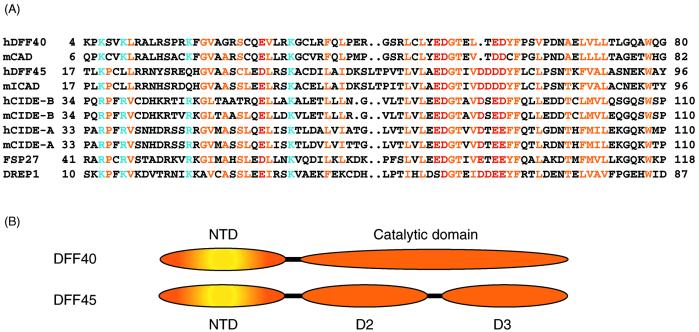
(A) Sequence alignment of CIDE proteins. Residues conserved in the family are colored yellow for hydrophobic residues, blue for basic residues, and red for acidic residues. (B) Domain structures (NTD and the catalytic domain) of DFF40 and (NTD, D2, and D3) of DFF45 are shown schematically.

