Figure 1.
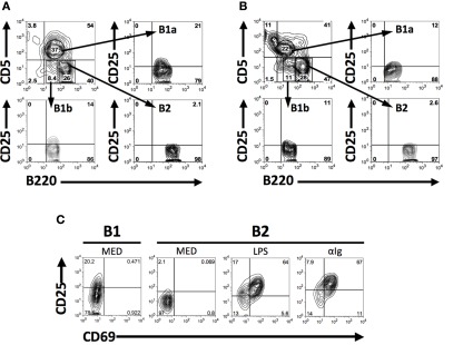
CD25 is expressed on a subset of naïve peritoneal B1 cells. (A,B) Freshly isolated BALB/c (A) and C57BL/6 (B) peritoneal washout cells were immunofluorescently stained for surface expression of B220, CD5, and CD25. Gates were set to identify B1a (B220lo, CD5+), B1b (B220lo, CD5−), and B2 (B220+, CD5−) cells among B220+ B cells and expression of CD25 was assessed for each population. Representative results from one of seven (A) and three (B) comparable experiments are shown. (C) Freshly isolated BALB/c B220loCD5+ B1a cells, and BALB/c splenic B220+CD5− B2 cells cultured in medium (MED) or stimulated with LPS (25 μg/ml) or with F(ab′)2 fragments of goat anti-mouse IgM (15 μg/ml) for 2 days, were immunofluorescently stained for CD25 and CD69. CD25 mean fluorescence intensity values (above background isotype staining) for CD25+ B1a cells and for CD25+CD69+B2 cells after stimulation with LPS and anti-Ig were, respectively, 53, 123, and 189. One of three comparable experiments is shown.

