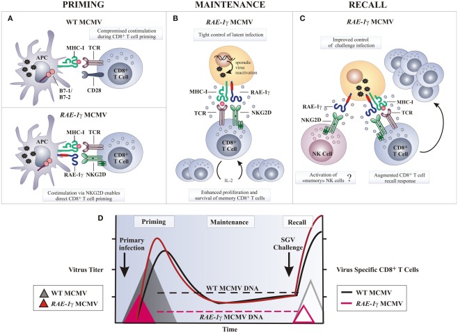
Figure 4.
Potential mechanisms of amplified CD8 T cell response following RAE-1γMCMV infection. (A) Naive CD8 T cells require engagement of the TCR by MHC-I molecules presenting antigen along with co-stimulatory signals, such as CD28 ligation by molecules of B7 family, to be fully activated. Wild-type MCMV (WT MCMV) dowmodulates B7.1 and B7.2 from the surface of infected APCs resulting in compromised T cell priming. Following RAE-1γMCMV infection co-stimulation via RAE-1γ–NKG2D interaction may replace B7-CD28 interaction and allow optimal CD8 T cell priming. (B) Stochastic MCMV reactivation during latency enables memory CD8 T cells co-stimulation via NKG2D–RAE-1γ interaction. Consequently, memory CD8 T cells are maintained or even inflated over time despite significantly reduced viral DNA load. (C) Challenge infection or any other serious stress leads to reactivation of latent RAE-1γMCMV and enhanced virus specific T cell response as a consequence of NKG2D ligand expression on cells undergoing virus reactivation. (D) Despite profoundly attenuated replication in vivo and reduced latent viral DNA load as compared to the WT MCMV, RAE-1γMCMV efficiently primes and maintains virus specific CD8 T cell response. In a challenge infection, mice immunized with the RAE-1γMCMV (red line triangle) resist salivary gland virus (SGV) challenge better as compared to mice initially infected with the WT MCMV (gray line triangle). APC, antigen presenting cell.

