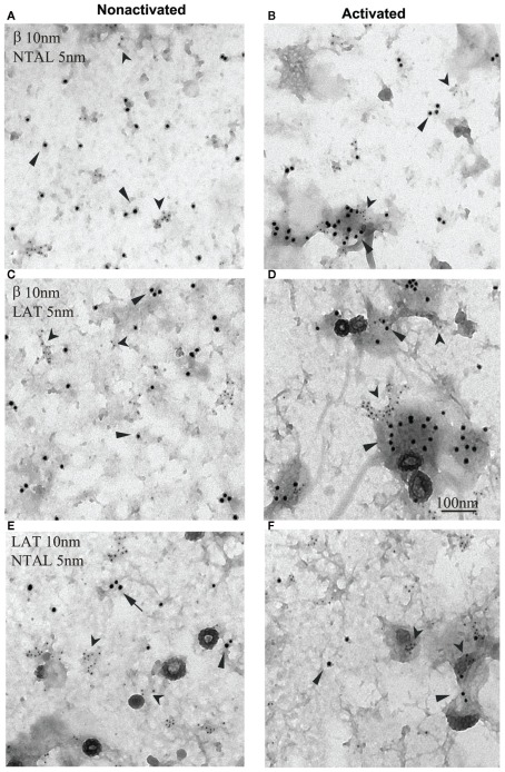
Figure 3.
Non-T cell activation linker, LAT, and FcεRI occupy different membrane microdomains in non-activated and FcεRI-activated cells. Plasma membrane sheets were isolated from non-activated (A,C,E) or antigen-activated (B,D,F) RBL-2H3 cells and double-labeled from the cytoplasmic side with first layer antibodies specific for the target structures and second layer anti-antibodies labeled with 5 or 10 nm gold particles. (A,B) FcεRI β subunit labeled with 10 nm gold (arrows) and NTAL labeled with 5 nm gold (arrowheads). (C,D) FcεRI β subunit labeled with 10 nm gold (arrows) and LAT labeled with 5 nm gold (arrowheads). (E,F) LAT labeled with 10 nm gold (arrows) and NTAL labeled with 5 nm gold (arrowheads). Scale bar in (D), 100 nm. Originally published in Journal of Experimental Medicine (Volná et al., 2004) where all experimental procedure details have been described.

