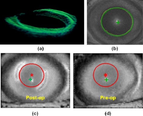
Fig. 5.
(a) Segmented (green). (b) Pupil edge fitting (detected edge in yellow and ellipse fitting in green). (c) ICRS inner diameter edge fit in the post-operative cornea (red line, and center as a red asterisk) and pupil center (green cross) (d) Evaluation of the same optical zone (in red) in the pre-operative cornea, using the pupil center as a reference.

