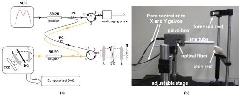
Fig. 1.
(a) Schematic of the SD-OCT system. M: mirror, L: lens, DC: dispersion compensation unit, PC: polarization controllers, DG: diffraction grating, C: circulators (b) Stage for human oral imaging, comprised of the custom-made probe and a head frame. For clarity, the fibers attached to the input (80/20) and output (50/50) couplers are shown in two different colors.

