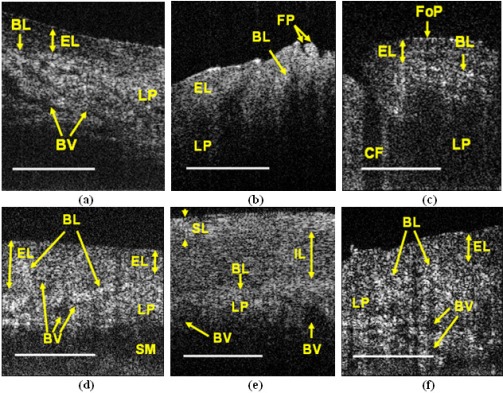
Fig. 4.
In vivo OCT images of human oral tissue, (a) labial; (b) dorsal surface of the tongue; (c) lateral surface of the tongue; (d) ventral surface of the tongue; (e) buccal; (f) soft palate. SL: superficial layer, IL: intermediate layer, EL: epithelial layer, BL: basal layer, LP: lamina proria, BV: blood vessel, FP: filiform lingual papillae, FoP: foliate lingual papillae, CF: circular furrow, SM: submucosa. Scale bar = 500 μm

