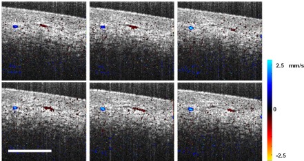
Fig. 6.
Consecutive Doppler images and Doppler OCT multimedia of the microvessels in the human labial tissue. The color bar represents the velocity of scatterers in the axial direction. Blue and red hues correspond to two opposite blood flow directions. Microvasculature as small as 16 μm is detectable using the SD-OCT system. Scale bar = 500 μm. Lateral field of view in the multimedia: 1 mm by 1 mm (Media 1 (4.2MB, MPG) )

