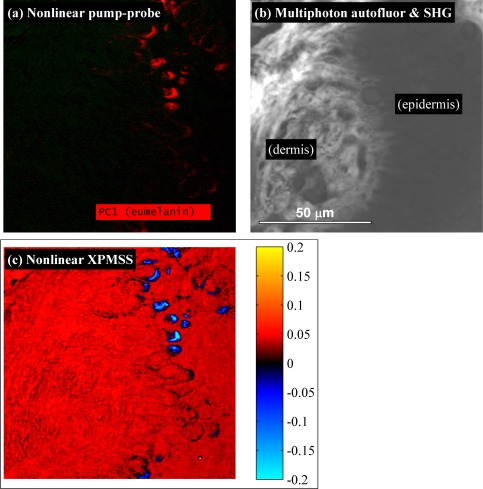
Fig. 5.
Images of the dermo-epidermal junction in a melanoma biopsy. a) Pump-probe image showing uniform eumelanin content. b) Combined multiphoton autofluorescence and SHG (imaged with a single PMT). c) XPMSS dot product image. Images acquired with a 40x 0.8 NA water immersion objective, 5 mW 720 nm pump, 5 mW 810 nm probe, 49 delays, 4 frame averaging.

