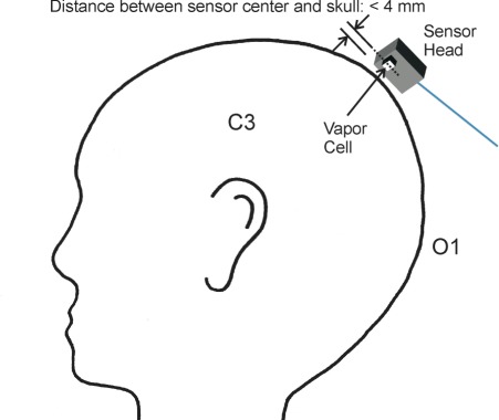
Fig. 3.
Sketch of the measurement positions on the head used to detect magnetoencephalographic signals. Spontaneous activity around 10 Hz linked to closing and opening of the eyes was measured with the sensor positioned above O1 (international 10-20 system for electrode positioning), whereas signals related to an electrical stimulation at the wrist were obtained over position C3.

