
Figure 1.
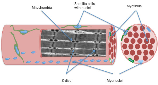
A schematic drawing of a muscle fiber (muscle cell) in longitudinal- and cross sectional plane. The muscle fiber is surrounded by two membranes, the plasma membrane (inner) and the basal lamina (outer). The satellite cells are located between these two membranes, and just beneath the plasma membrane lays the myonuclei. The contractile proteins in the muscle cell are arranged in myofibrils. In the longitudinal plane you see that the myofibrils are organized into sarcomeres separated by the z-disc and the mitochondria are seen as circular spots between the myofibrils.