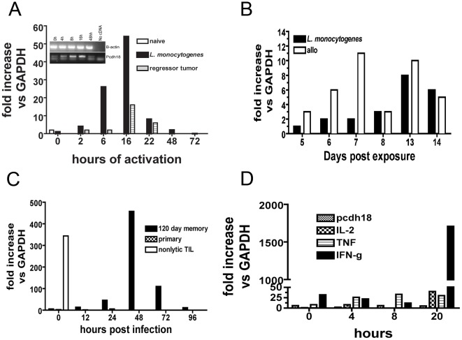
Figure 3. RNA analyses of spleen cells after induction of memory in vivo.
(a) qRT-PCR analysis of spleen CD8+ T cells isolated from BL/6 mice previously infected with either 5,000 recombinant Listeria monocytogenes, buffer controls, or EL4 cells at a subtumorigenic dose (‘regressor tumor’). 28 days after exposure spleen CD8+ T cells were purified, activated in vitro with Con A for the indicated times, RNA prepared and qRT-PCR performed as described in ‘Materials and Methods’. (This analysis has been performed using mice previously infected for times up to 1 year with similar results). Insert shows gel analysis of pcdh18 RT-PCR expression from L. monocytogenes-immune mice. (3b) qRT-PCR analysis of purified CD8+ T cells from mice injected with L. monocytogenes or allogeneic H-2D spleen cells. (3c) qRT-PCR analysis of purified CD8+ T cells from mice originally infected with L. monocytogenes which were challenged by in vivo infection by L. monocytogenes. Spleens were isolated at the indicated times following challenge. Age-matched naive mice received only primary exposure given at the time of secondary challenge. Nonlytic TIL are shown for comparison. (3d) qRT-PCR analysis of purified control CD8+ spleen T cells and activated in vitro with anti-CD3e for the indicated times before RNA isolation and analysis by qRT-PCR. The data shown are representative of multiple repetitions.

