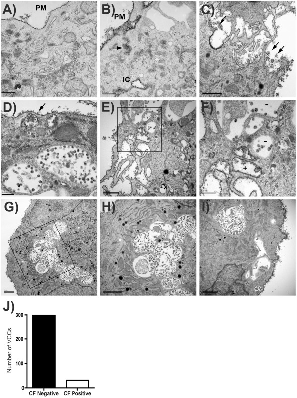
Figure 7. The majority of virus-containing compartments in infected MDMs are inaccessible to a cell surface label.
(A-B) Uninfected Human MDMs cultured on ACLAR embedding film were fixed and stained with cationized ferritin (CF), followed by standard electron microscope processing procedures. Images were then obtained under a Hitachi H-7500 transmission electron microscope. Cationized ferritin labeled plasma membrane is seen along the plasma membrane (PM). (A) and (B) represent uninfected macrophages, bars = 0.5 µm. IC = apparent intracellular space stained with CF. (C) HIV particles were seen at PM and stained with CF on periphery of cells (arrows). Bar = 1 µm. (D) CF is seen staining HIV particles underlying a PM fold (arrow), while deeper VCCs lack CF staining. Bar = 0.5 µm. (E-F) CF staining of membrane protrusions contrasts with lack of CF in intracellular VCCs (F is higher magnification view of boxed region in E). Bar = 1.0 µm (E), 0.5 µm (F). (G-I) Additional views of PM staining with CF and exclusion of CF from VCC. (H represents higher magnification view of boxed region from G, bars = 2 µm). (J) VCCs were counted as CF+ or CF- from 329 apparent intracellular VCCs in more than 50 cells. The number of CF-negative compartments vs. CF-positive compartments is indicated.

