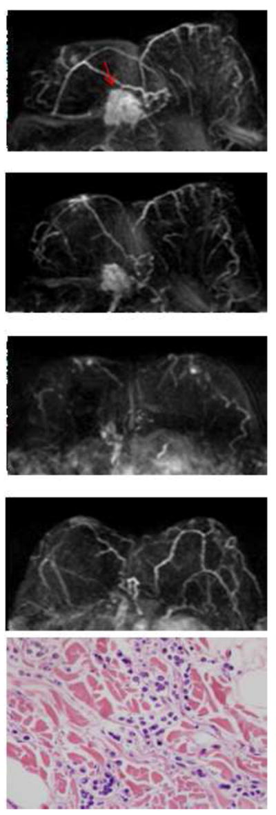
Figure 4.

A hormonally positive, HER2 negative infiltrating lobular cancer in the right breast. The first image is baseline MRI prior to NAC. The second and the third images are follow-up MRI during NAC treatment. The fourth image is the last MRI after the completeness of NAC treatment. Following NAC, MRI showed a 0.5cm residual tumor, which differs markedly from a 6.3 cm tumor found in surgical pathology. In pathology, scattered residual tumor cells surrounded by chemotherapy associated changes, which include fibrosis and a lymphocyte infiltrate, are noted.
