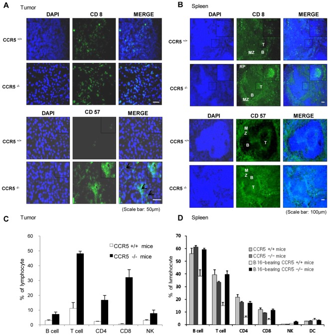
Figure 5. Induction of the infiltration of CD8+ T cells and NK cells into tumor, and increase in spleen of CCR5−/− mice.
A and B, Immunolfluorescence analysis was used to determine the expression levels of CD8 (CD8+ cytotoxic T-cell surface marker) and CD57 (NK cell marker) in tumor and spleen sections. The images shown are representative of three separate experiments performed in triplicate. Scale bars indicate 50 µm (A) and 100 µm (B). C and D, Analysis of lymphocyte phenotypes. The tumor and spleen tissues were separated at study termination (Day 31). Flow cytometry analysis was performed using FACSAria flow cytometry, and represent data were shown. Data are means ± S.D. of four experimental animals.

