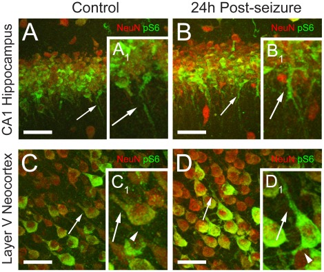
Figure 5. Phospho-S6 (Ser235/236) expression is highly upregulated in the cell bodies and dendrites of hippocampal and neocortical pyramidal neurons at 24 h following neonatal seizures.
A-B. Confocal images of CA1 hippocampal region double labeled with NeuN (red) and phospho-S6 (green) show a moderate increase in phospho-S6 expression in pyramidal neuron cell bodies and a marked phospho-S6 upregulation in the dendrites (arrows) in the post-HS group (B, B1), relative to controls (A, A1). C-D. Confocal microscopy of neocortical layer V demonstrates a similar predominant increase in phospho-S6 (green) expression in the apical (arrows) and basal (arrowheads) dendrites of NeuN (red) positive neurons in the HS group (D, D1), relative to controls (C, C1). Confocal images represent z-stacks composed of multiple plane images collected at 1 µm intervals. Scale bars are 100 µm for A-D. Insets (A1–D1) show corresponding higher magnification of individual cells.

