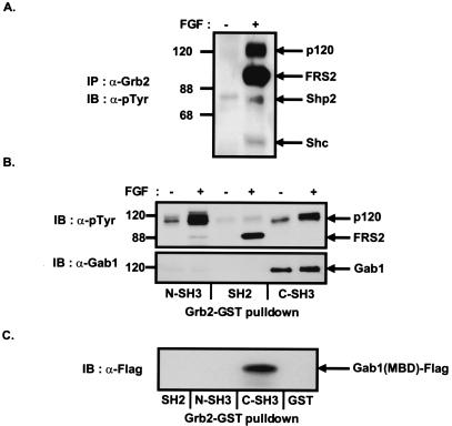
Figure 1.
Complex formation between Grb2 and Gab1 in FGF-stimulated cells. (A) Quiescent Swiss 3T3 cells were unstimulated or stimulated with FGF1 and heparin (100 ng/ml and 5 μg/ml, respectively) for 10 min. Lysates were immunoprecipitated with anti-Grb2 antibodies and followed by SDS–PAGE and immunoblotting with anti-pTyr antibodies. (B) Swiss 3T3 cells were unstimulated or stimulated with FGF1 and heparin (100 ng/ml and 5 μg/ml, respectively) for 10 min. Cell lysates were incubated with immobilized GST fusion proteins of the N-terminal SH3, SH2, or C-terminal SH3 domains of Grb2. Bound proteins were eluted and resolved by SDS–PAGE and then followed by immunoblotting with anti-pTyr (Upper) or anti-Gab1 antibodies (Lower). (C) Gab1 binds to the C-terminal SH3 domain of Grb2 through the MBD. 293 cells were transfected with the expression vector for Flag-tagged MBD of Gab1. The lysates were incubated with immobilized GST fusion proteins of the SH2, N-SH3, or C-SH3 domains of Grb2. Bound proteins were eluted and resolved by SDS–PAGE and followed by immunoblotting with anti-Flag antibodies.

