Figure 6.
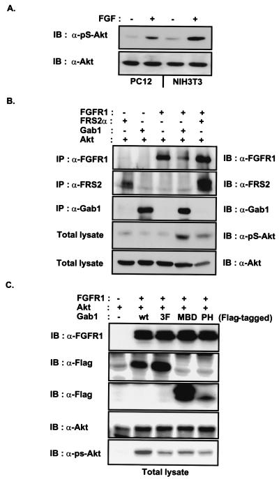
FRS2 and Gab1 potentiate FGF-induced activation of Akt. (A) Quiescent PC12 or NIH 3T3 cells were unstimulated or stimulated with FGF1 and heparin (100 ng/ml and 5 μg/ml, respectively) for 5 min. Equivalent amounts of total cell lysates were resolved by SDS–PAGE and then followed by immunoblotting with anti-pS-Akt or anti-Akt antibodies. (B) The 293 cells were transfected with expression vectors for FGFR1, FRS2α, Gab1 and Akt as indicated. The expression of FGFR1, FRS2α, and Gab1 was verified by immunoprecipitation with antibodies specific to these proteins and followed by SDS–PAGE and immunoblotting with the same antibodies. Equivalent amounts of total cell lysates were resolved by SDS–PAGE and immunoblotted with anti-pS-Akt or anti-Akt antibodies. (C) The MBD of Gab1 inhibits Gab1-potentiated FGFR-induced activation of Akt. 293 cells were transfected with the expression vectors for FGFR1, Akt and Gab1-Flag, Gab1(3F)-Flag, Gab1-MBD-Flag, or Gab1-PH-Flag as indicated. Equivalent amounts of total cell lysate were resolved by SDS–PAGE and followed by immunoblotting with anti-FGFR1, anti-Flag, anti-Akt, or anti-pS-Akt antibodies.

