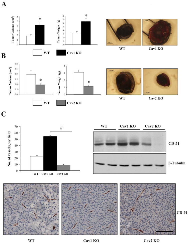
Figure 1. Cav1 ablation in mice promotes the growth of B16F10 melanoma cells independently of Cav2.

106 B16F10 melanoma cells were orthotopically (intradermally) implanted in the skin of 3-4-month-old WT, Cav-1 KO (A) and Cav2KO (B) C57Bl/6 female mice (n≥8, per group). After 18 days, tumors were excised and their size determined. Representative images of tumors are displayed on the right. C, CD31 immunohistochemistry of tumor sections showing that microvascular density correlates with tumor size in Cav1KO and Cav2KO mice (n=5, per group). CD31 immunoblotting of whole-tumor lysates is shown. Results are means ± SEM shown (* #, P < 0.05, by two tailed Mann-Whitney and by Dunnett’s Multiple Comparisons Test; Scale Bar, 100μm).
