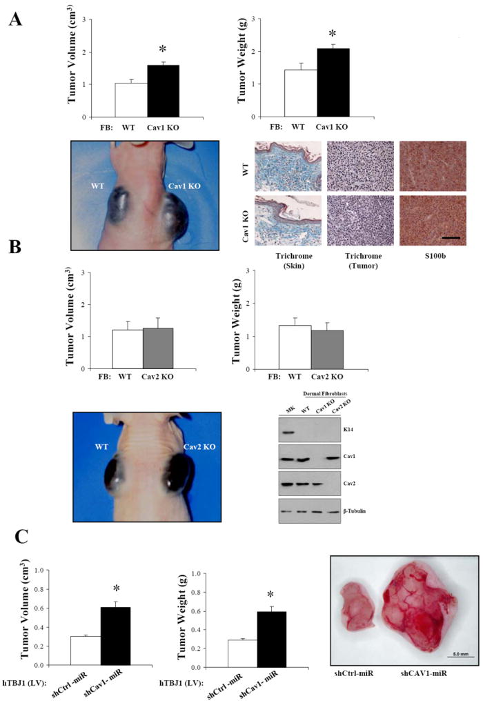
Figure 2. Absence of Cav1 but not Cav2 in dermal fibroblasts enhances the growth of melanoma cells in co-injection experiments.

A, B16F10 melanoma cells (1×10⁵) were intradermally co-injected with Cav1KO neonatal dermal fibroblasts at 1:5 ratios in 3-4-month-old nude female mice. After 14 days, tumors were dissected and their size determined. Masson’s trichrome staining and S100b (melanoma cell marker) immunohistochemistry of tumor sections revealed similar intratumoral collagen deposition between WT-FB/B16F10 and Cav1KO FB/B16F10 xenografts (n=5, per group). B, tumor size of B16F10 cells coinjected with WT and Cav2KO dermal fibroblasts as in (A). K14 (keratinocyte marker), Cav1 and Cav2 immunoblots of freshly isolated dermal fibroblast and keratinocytes (MK) are also shown. β-Tubulin immunoblot is shown as loading control. C, lentivirus mediated CAV1 silencing (Lv-shCAV1miR) in hTBJ cells promotes the growth of A-375 cells as determined by co-injection experiments. Results are means ± SEM (n≥5, per group; *, P < 0.05, by two tailed Mann-Whitney Test; Scale Bar, 100μm).
