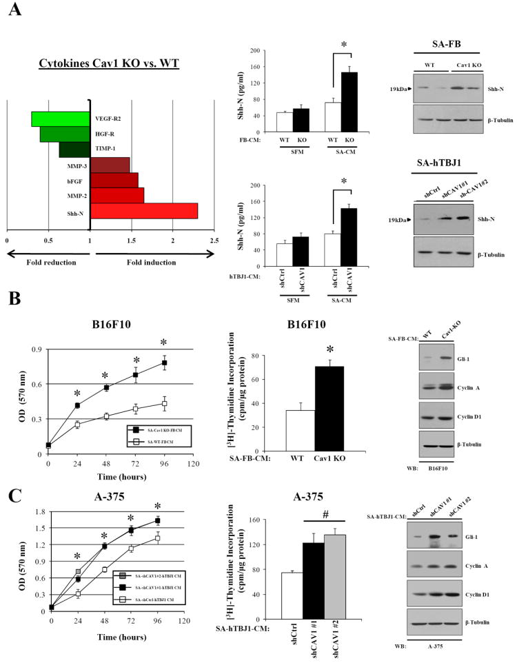
Figure 4. Serum-activated Cav1KO dermal fibroblasts display increased amounts of protumorigenic cytokines.

A, left, cytokines differentially regulated in conditioned medium from SA-WT and SA-Cav1KO neonatal dermal fibroblasts. Center, ELISA showing increased ShhN levels in the conditioned medium of serum-activated dermal fibroblasts (SA-CM) lacking Cav1 (n= 4 per group). ShhN immunoblots of serum-activated dermal fibroblasts are displayed (right). B, MTT-assay and [3H]-Thymidine incorporation assay (48h) of B16F10 melanoma cells treated with SA-CM from WT and Cav1KO fibroblasts. Immunoblot analysis showing increased expression of Gli-1, Cyclin D1 and Cyclin A in B16F10 melanoma cells incubated (48h) with SA-CM from Cav1KO dermal fibroblasts (n= 8 per group). Similar results are shown (C) for human A-375 melanoma cells treated with SA-CM from hTBJ1-shCtrl and hTBJ1-shCAV1 cells. Results are means ± SEM (n ≥ 6 per group; *, #, P < 0.05, by two tailed Student’s t test and by Dunnett’s multiple comparisons test).
