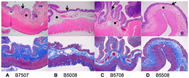
Figure 3. Colon H&E and collagen deposition.

H&E staining (top) and collagen staining (Trichrome, bottom) of the colon. B7507 (A), B5508 (B), B5008 (C) and B5708 (D) (magnification ×4). A moderate to extensive increase in collagen can be seen within the submucosa (asterix, top figures) and is illustrated by blue staining by Trichrome (bottom figures). Mild to extensive collagen deposition is also present between the glands within mucosa (arrow). The most severe changes are illustrated in B7507 with a thick layer of dense submucosal fibrous connective tissue and regions of complete mucosal epithelial cell loss secondary to collagen deposition.
