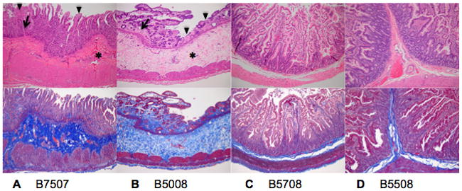
Figure 4. Small intestine H&E and collagen deposition.

H&E staining (top) and collagen staining (Trichrome, bottom) of the small intestine of B7507 (A), B5508 (B), B5008 (C) and B5708 (D) (magnification ×10). A marked increase in collagen is found in both the lamina propria (arrow) and submucosa (asterix) of B7507 and B5508 in addition to moderate (B7507) to severe (B5508) blunting and loss of villi with necrosis and loss of surface enterocytes (arrowhead). Mild villous blunting is present in B5508. The small intestine of B5008 and B5708is has a normal appearance.
