
Fig. 1. Anti-inflammatory role of MUC1 during airway infection.
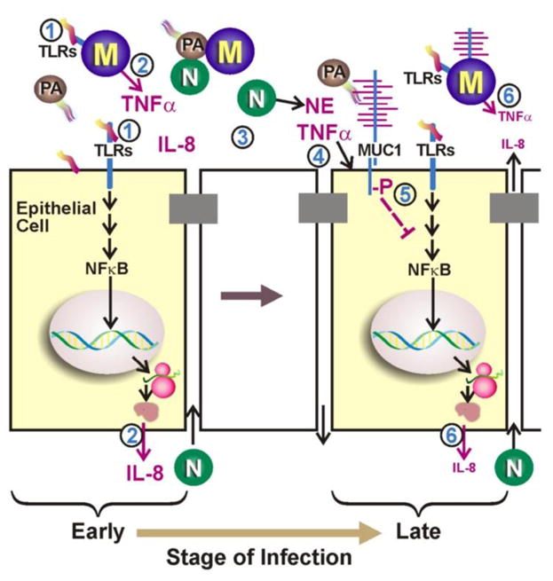
(Step 1) During the early stage of infection by P. aeruginosa (PA), bacterial PAMPs (e.g. flagellin) activate TLRs and NF-κB on epithelial cells and macrophages (M). (2) Activation of NF-κB leads to increased expression of TNF-α and of IL-8, which are subsequently secreted. (3) IL-8 recruits neutrophils (N) across the epithelial barrier that release NE into the lumen of the airways. (4) NE and TNF-α up-regulate MUC1 gene expression resulting in increased expression of MUC1 mucin at the apical surface of lung epithelial cells. (5) During the late stage of infection, tyrosine phosphorylation of MUC1 CT domain leads to inhibition of TLR signaling and (6) down-regulation of inflammation. (Fom Kim and Lillejoj [36])