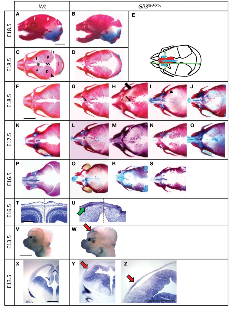
Figure 1.
Frontal bone development in Gli3Xt-J/Xt-J mice. Alcian blue and Alizarin red S stained heads of Wt (A,C,F,K,P) and Gli3Xt-J/Xt-J (B,D,G–J,L–O,Q–S) embryos and a schematic of Gli3Xt-J/Xt-J head indicating heterotopic ossification (red), heterotopic cartilage formation (blue), and measurements of the head (green arrows) (E). At E18.5 Gli3Xt-J/Xt-J frontal bones are abnormally shaped (black arrow in H) and in the interfrontal suture there are heterotopic bones [(G,H); double-lined arrow] that have fused in some samples causing suture synostosis. In samples with less heterotopic bones the suture is wider compared to Wt [(I); arrowhead]. At E17.5 Gli3Xt-J/Xt-J frontal bones have similar features as at E18.5. At E16.5 Gli3Xt-J/Xt-J frontal bones are hypoplastic, but ectopic ossification is already evident. Ectopic cartilage is seen in the interfrontal suture of Gli3Xt-J/Xt-J mice [(I,J,L,N,O,Q,R,U); green arrow]. Toluidine blue stained frontal sections through the posterior interfrontal suture of Wt (T) and Gli3Xt-J/Xt-J (U) heads at E16.5. Morphology of the Gli3Xt-J/Xt-J brain is abnormal (U). Dash line in T and U indicate the midline of the head. Alkaline phosphatase stained E13.5 heads of Wt (V,X) and Gli3Xt-J/Xt-J (W,Y,Z) embryos, where heterotopic osteoblast differentiation is detected in Gli3Xt-J/Xt-J mice [(W,Y,Z); red arrow]. f, Frontal bone; i, interparietal bone; is, interfrontal suture; ls, lambdoid suture; p, parietal bone; ss, sagittal suture. Scale bars: 2 mm, except 300 μm in (T) and 500 μm in (X,Z).

