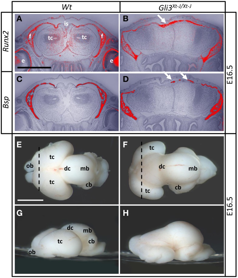
Figure 2.
Osteoblast differentiation and brain morphology of Gli3Xt-J/Xt-J mice at E16.5. Frontal sections of Wt (A,C) and Gli3Xt-J/Xt-J heads (B,D). (A–D) The osteoblastic markers Runx2 and Bsp are expressed in the frontal bones and ectopically in the interfrontal suture (arrows) of Gli3Xt-J/Xt-J mice. Dash line in (E,F) represent the plane of section in (A–D) respectively. Brains of Wt (E,G) and Gli3Xt-J/Xt-J (F,H) mice. The Gli3Xt-J/Xt-J brain has hypoplastic dorsal telencephalon and diencephalon resides more anteriorly. The cerebellum extends more ventrally. cb, Cerebellum; dc, diencephalon, f, frontal; is, interfrontal suture; e, eye; mb, midbrain; ob, olfactory bulb; tc, telencephalon. Scale bars are 1 mm in (A), 2 mm in (E).

