
Figure 1.
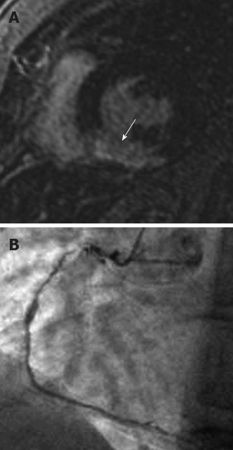
A 42-year-old female who presented with acute chest pain to the emergency department. She had no risk factors for coronary artery disease and the clinical suspicion was of myocarditis. A: Late-enhancement short axis sequence shows a transmural area (arrow) of high signal involving the inferior segment consistent with an acute myocardial infarction involving the right coronary artery territory. Note that it is an acute rather than chronic infarct, because there is no wall thinning; B: An invasive angiogram confirmed diffuse coronary artery disease throughout the right coronary artery. Note that the likelihood of recovery of this segment with revascularization is extremely low because it is a transmural infarct.