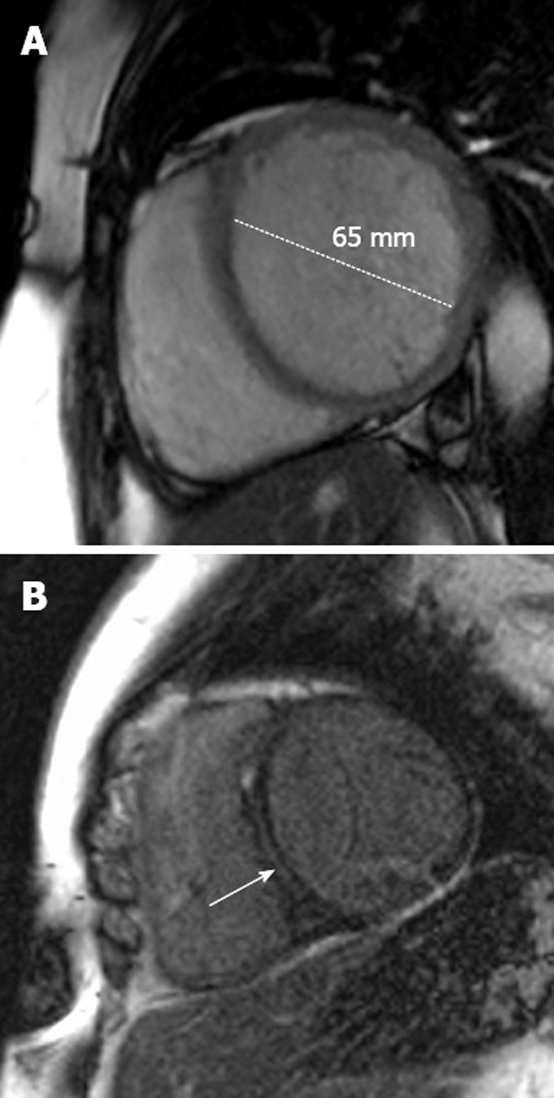
Figure 3.

Dilated cardiomyopathy. A: A 48-year-old man with progressive shortness of breath. The short-axis steady-state free precession sequence demonstrates a dilated left ventricle with a thin wall characteristic of dilated cardiomyopathy (DCM); B: Late-enhancement short-axis image shows late-enhancement in the interventricular septum (arrow). This is a characteristic location for fibrosis detection in idiopathic DCM and effectively excludes an ischemic etiology. Such late-enhancement has prognostic implications for patients with idiopathic DCM, being associated with an increased prevalence of all-cause death, hospitalization, sudden cardiac death and ventricular tachycardia.
