Figure 2.

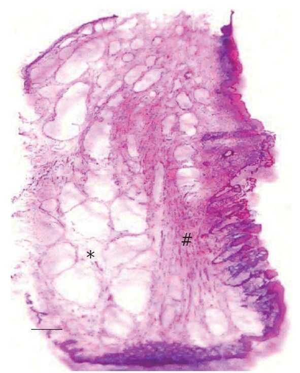
Pathological changes in hemorrhoids. *: Marked dilatation of hemorrhoidal venous plexus; #: Fragmented anal subepithelial muscle (the Treitz’s muscle or mucosal suspensory ligament) (Scale bar = 1 mm).

Pathological changes in hemorrhoids. *: Marked dilatation of hemorrhoidal venous plexus; #: Fragmented anal subepithelial muscle (the Treitz’s muscle or mucosal suspensory ligament) (Scale bar = 1 mm).