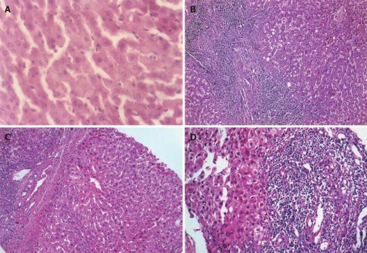
Figure 1.
Hematoxylin and eosin staining of liver tissues (HE, × 200). A: Control group showed normal lobular architecture and cell structure; B: Patients before treatment showed fibro-proliferated bile ductules, thick fibrous septa and dense inflammatory cellular infiltration; C: The conventional group showed moderate fibrosis with inflammatory infiltration and slight ballooning of liver cells; D: Aloe vera high molecular weight fractions group showed minimal infiltration and minimal fibrosis.

