
Figure 4.
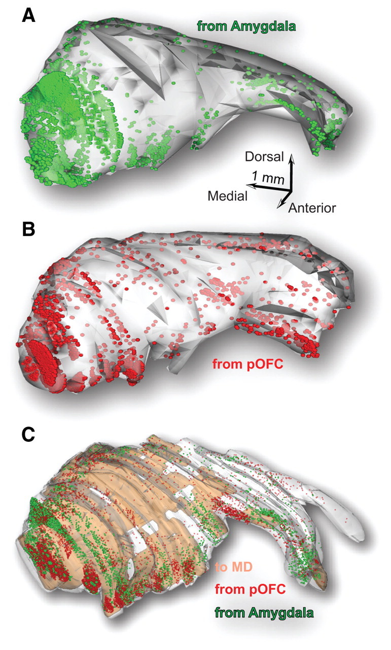
Map of TRN reconstructed in 3D shows terminations from the amygdala, pOFC, and MD. A, B, Serial sections were used to reconstruct the TRN in three dimensions and map terminals from the amygdala (green; A) and the pOFC (red; B) in each case. C, The relative coordinates of these pathways were used to superimpose all plotted terminals from the amygdala and pOFC and the TRN neurons that project to MD (brown shading) onto a reference 3D model of the TRN. The 3D map shows the widespread distribution of labeling and sites of high or low overlap.