
Figure 5.
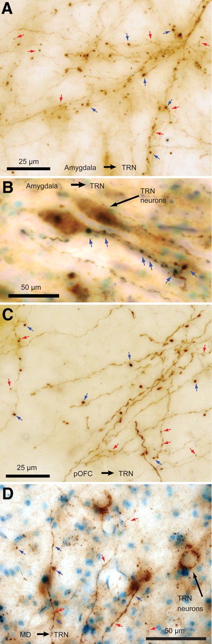
Labeled pathways from the amygdala, pOFC, and MD terminated with large and small boutons in the TRN. A, B, BDA-labeled fibers from the amygdala with large (blue arrows) and small (red arrows) boutons. Two TRN neurons are visible in B (long black arrow), surrounded by large axon terminals from the amygdala closely apposed to the cell bodies and proximal dendrites. C, Labeled fibers from pOFC. D, Labeled fibers from MD in the TRN.