
Figure 7.
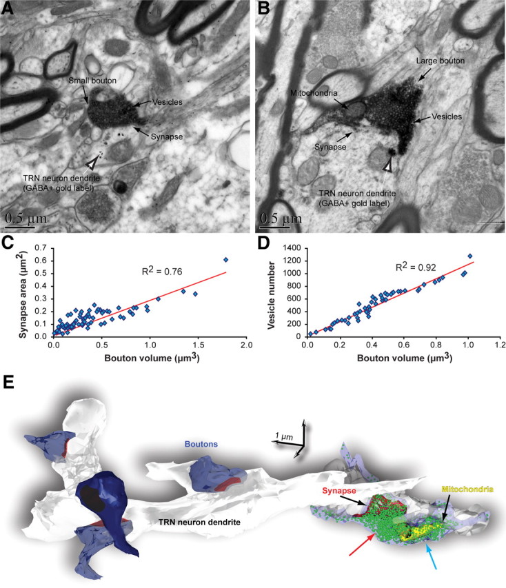
Synaptic features of labeled boutons from the amygdala in TRN. A, B, EM images show small (A) and large (B) labeled boutons (dark flocculent DAB staining; black arrows) forming asymmetric (presumed excitatory) synapses with dendrites of GABAergic TRN neurons (labeled with gold particles; black and white arrows). C, D, Bouton volume was positively correlated with synapse area (C) and vesicle number (D). E, 3D reconstruction of some labeled boutons from the amygdala (blue) that form synapses (red) with a dendrite from an inhibitory TRN neuron (white/gray). A large bouton on the right was rendered transparent to show the position of vesicles (green spheres) and mitochondria (yellow). In large boutons, vesicles were aggregated into two clusters, one found adjacent to the postsynaptic density (red arrow) and another positioned distal to the postsynaptic density (blue arrow).