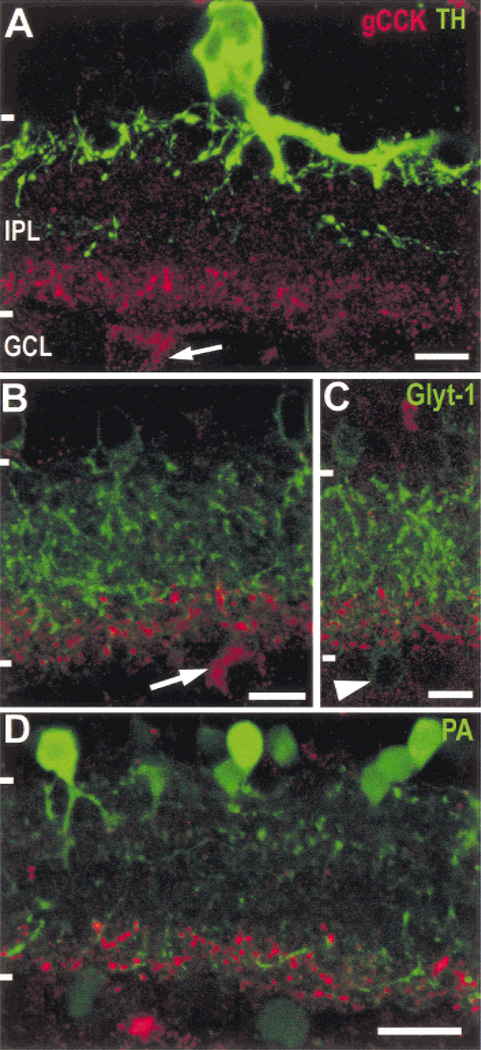
Fig. 2. Gastrin CCK-IR amacrine cells do not contain tyrosine hydroxylase (TH) or glycine transporter-1 (Glyt-1), and the gCCK-IR varicosities costratify with parvalbumin immunoreactivity.
A: The TH-IR (green) processes are dense in the outer stratum of the IPL; there is also a sparse plexus of TH-IR processes in the center of the IPL. The large TH-IR amacrine cell soma is seen in the typical position in the inner row of somata in the INL. A gCCK-IR (red) amacrine cell soma (arrow) is shown in the GCL in peripheral retina. Neither the varicose gCCK-IR processes nor the perikarya contain TH-IR. Stack = 13 × 0.5 µm. B,C: Glyt-1 immunoreactivity (green) is present in processes throughout the IPL, as well as in a population of amacrine cells in the INL. The gCCK-IR (red) processes are associated with some of the Glyt-1 processes in the inner stratum of the IPL. Neither the perikaryon (B, arrow) nor the gCCK-IR processes contain Glyt-1 immunoreactivity. Glyt-1-IR perikaryon (C, arrowhead) in the ganglion cell layer did not contain gCCK immunoreactivity. Stack = 3 × 0.5 µm (B) and 7 × 0.5 µm (C). D: The parvalbumin-IR (green) perikarya in the inner nuclear layer (INL) are mostly AII-like amacrine cells, with dendrites in the inner and outer strata of the inner plexiform layer (IPL). There are also fainter parvalbumin-IR somata in the ganglion cell layer (GCL). Many of the gCCK-IR (red) varicosities are found in the vicinity of parvalbumin-IR processes in the inner stratum of the IPL. Stack = 5 × 0.5 µm. Scale bars = 10 µm (A, B, & C) and 20 µm (D).

