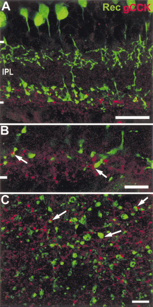
Fig. 6. Gastrin CCK-IR processes contact ON-cone bipolar cell axon terminals.
A: Type 2 and 8 cone bipolar cells are labeled with antisera to recoverin (green) in a vertical section. The ON-cone bipolar cells (type 8) have axon terminals in the vicinity of the gCCK-IR (red) varicosities in stratum 5. Stack = 7 × 0.5 µm. B: Contacts between the gCCK-IR varicosities and recoverin-IR ON-cone bipolar cells are seen at a higher magnification of a vertical section (arrows). Single optical section = 0.5 µm. C: The contacts (arrows) between gCCK-IR varicosities and recoverin-IR ON-cone bipolar cell terminal are seen more clearly in a single optical section (0.5 µm) in stratum 5 of the IPL. Scale bars=25 µm (A) and 10 µm (B & C).

