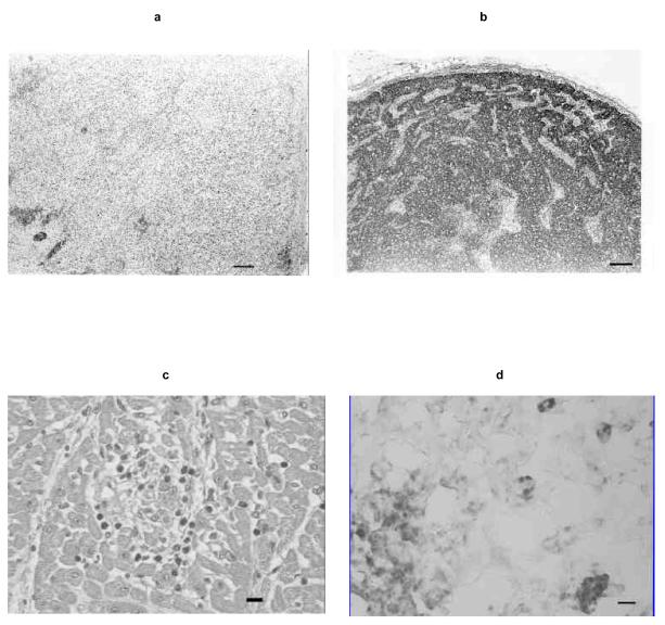
Fig. 5.
(a) Photomicrograph of a SCID2 lymph node section, lacking follicles and lymphocytes. Bar, 100 μm. (b) Photomicrograph of a SCID1 lymph node section, fully populated with lymphocytes. Bar, 100 μm. (c) Photomicrograph of a SCID1 heart section containing a focus of myocardial necrosis with lymphocyte infiltrates. Bar, 10μm. (d) Photomicrograph of macrophages labeled by immunohistochemistry (dark-staining cells) in a SCID1 spleen section. Bar, 10μm.

