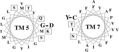
Figure 5.
Helical wheel representation of TMs 5 and 7 of LdNT1.1. Residues of TM 5 and TM 7 are shown on a helical wheel of 3.6 residues per turn. Amino acids with polar side chains are indicated within squares. The positions of the G183D (G → D) and C337Y (C → Y) mutations in TMs 5 and 7, respectively, are also indicated.

