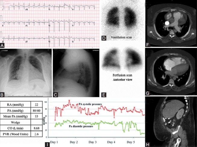
Figure 11.

Images obtained in 35-year-old male with cor pulmonale. (A) S1Q3T3 pattern noted consistent with right ventricle strain. RVH is evident from the prominent R wave in the early precordial leads. Evidence of pulmonary disease, with failure of R wave transition and presence of Rs in V4. (B, C) PA and lateral chest X-ray with severe 4-chamber cardiomegaly with pulmonary vascular redistribution and visible obesity. (D, E) Normal VQ scan, performed anteriorly due to excess obesity. (F-H) CT showing moderate to severe cardiomegaly with normal lung parenchyma, mild dependent hypoventilatory changes and abdominal obesity. (I) PA pressure measurements decrease over the course of first 5 days of hospitalization while receiving CPAP and intensive diuresis (red line: Systolic PAP, green line: Diastolic PAP).
