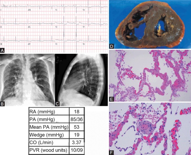
Figure 6.

Category 1 PH diagnosed postmortem as secondary to pulmonary capillary hemangiomatosis (PCH). (A) ECG shows mild left ventricular hypertrophy and right atrial enlargement. (B, C) Chest X-ray shows PA enlargement and cephalization with Kerly B lines. (D) Gross dissection of the heart shows thickened and dilated right ventricle. (E) Pulmonary capillary hemangiomatosis, seen as increased numbers of pulmonary capillaries swollen with large numbers of red blood cells, sharply demarcated from normal-appearing lung parenchyma (H and E, ×100). (F) Muscularization of a small-caliber arteriole. Surrounding alveolar septal capillaries with changes of pulmonary capillary hemangiomatosis (H and E, ×200).
