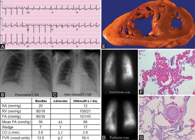
Figure 7.

Category 1 PH due to a second case of pulmonary capillary hemangiomatosis. (A) ECG showing sinus tachycardia and right atrial enlargement. (B) Chest X-ray on presentation with cardiomegaly and clear lung fields. (C) Chest X-ray after initiation of sildenafil showing pulmonary edema. (D) Ventilation-perfusion scan showing accentuation of basilar perfusion images. (E) Gross dissection of the heart shows thickened and dilated RV. (F) Patchy areas of pulmonary capillary proliferations engorged with red blood. (G) Concentric intimal fibrosis and medial hypertrophy of pulmonary arteries and arterioles.
