
Figure 3.
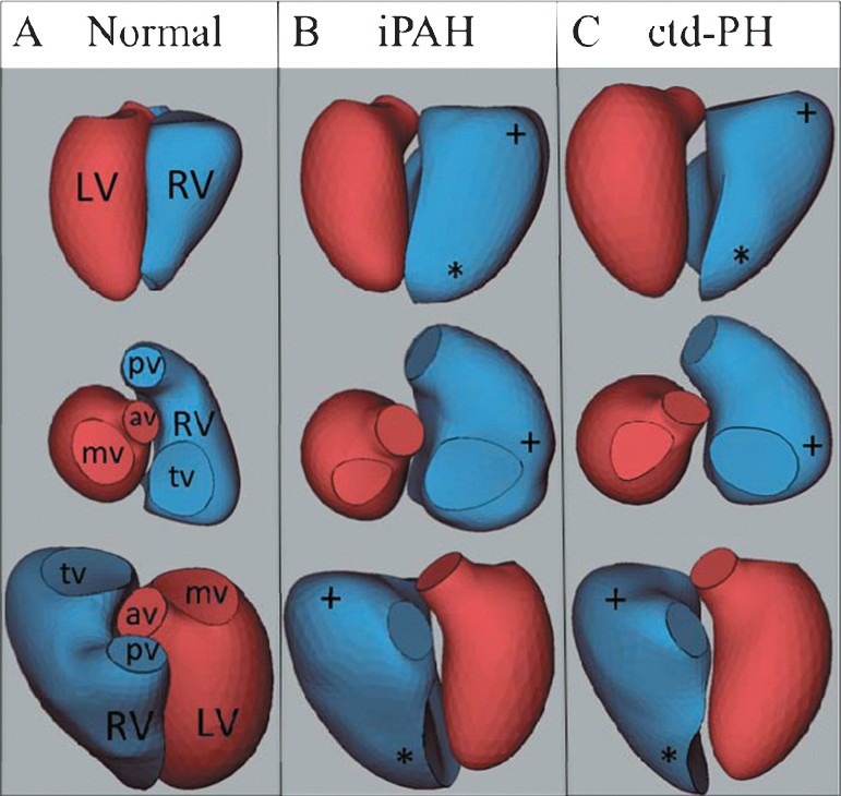
Representative 3D reconstructions from a normal (A), iPAH (B) and ctd-PH (C) heart demonstrating apical rounding (*) and basal bulging (+), [LV: Left ventricle; RV: Right ventricle, pv: Pulmonary valve, av: Aortic valve; tv: Tricuspid valve; mv: Mitral valve].