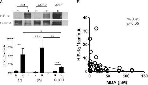
Figure 1.
HIF-1α protein in nuclei of peripheral blood mononuclear cells (PBMCs). PBMCs were incubated in hypoxic condition (1% oxygen, 5% CO2, and 94% N2) for 24 h. Nuclear protein was extracted, and HIF-1α protein was detected. A, HIF-1α protein was normalized by lamin A. Representative Western blot images are also shown. B, Correlation between HIF-1α protein in nuclei and sputum malondialdehyde (an oxidative stress marker). *P < .05; **P < .01; ***P < .001. H = hypoxia; HIF-1α = hypoxia inducible factor-1α; MDA = malondialdehyde; N = normoxia; NS = nonsmokers; SM = smokers without COPD.

