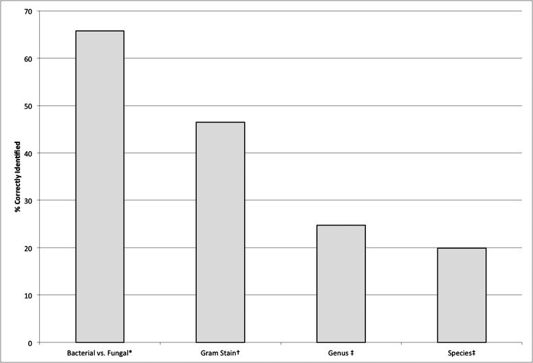
Figure 2.
The aggregate results when looking at all clinicians and applicable photographs. * Based on all photographs, n = 79 photos. † Gram stain was examined for bacterial ulcers only, n = 40 photos. ‡ Based on photographs of both fungal and bacterial ulcers with available microbiologic data. For genus, n = 70 photos. For species, n = 44 photos.

